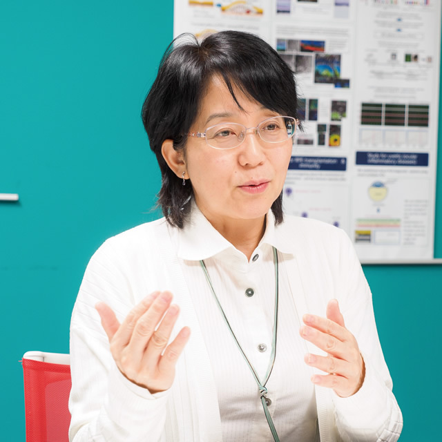
{}

Power of Innovation

iPS Cells Give New Hope Against Eye Diseases

Masayo Takahashi

Dr. Masayo Takahashi graduated from Kyoto University’s Faculty of Medicine in 1986. In 1992, she completed her Ph.D. in Visual Pathology at Kyoto University’s Graduate School of Medicine. She first worked as a clinician, but later became interested in research following her studies in the United States in 1995. In 2005, her lab became the first in the world to successfully differentiate neural retina from embryonic stem cells. She is currently the project leader of the Laboratory for Retinal Regeneration at the RIKEN Center for Developmental Biology (CDB).

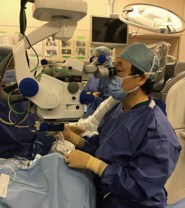
 In September 2014, a team led by Dr. Masayo Takahashi, Japan’s leading ophthalmologist, of the Japanese research institute RIKEN carried out the world’s first induced pluripotent stem cell (“iPS” cell) transplant surgery for an eye disease. The generation of iPS cells was first announced in 2006 by Professor Shinya Yamanaka, the director of Kyoto University’s Center for iPS Cell Research and Application (CiRA). These cells are capable of differentiating into various cell types that make up the tissues and organs in the body, and are believed to have potential uses in restoring cells lost by sickness or injury, bringing hope of providing cures to diseases for which there was previously no treatment. The iPS cell transplant surgery conducted by Dr. Takahashi and her team has garnered much attention as a remarkable achievement that is anticipated to lead to practical regenerative medical applications.
 
 “My interest in regenerative medical research began,” explains Dr. Takahashi, “when I accompanied my husband in his studies abroad in the United States and learned of the existence of stem cells that were capable of replenishing cells in the body. Although I was later successful using embryonic stem cells to produce retinal cells, I knew as soon as I heard about the discovery of iPS cells that I’d found the ideal stem cell. I credit our world’s-first achievement to being specialized in the clinical field of ophthalmology while also being exposed to stem cell research, which was a completely different field, and also to the fact that we’ve always kept pushing our research forward on the leading edge.”
 
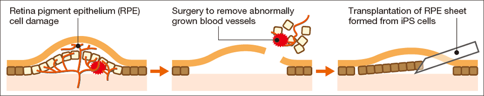
 In the above-mentioned surgery, iPS cells were made from the skin tissue of a patient who was suffering from wet-type age-related macular degeneration (AMD), and these iPS cells were differentiated into retinal pigment epithelial (RPE) cells and processed into the form of a cell sheet. The RPE cell sheet was then transplanted into the patient from whom the iPS cells were generated, which was able to prevent further deterioration of the patient’s eyesight (a symptom of wet-type AMD).

Method of the 2014 surgery

Wet-type age-related macular degeneration (AMD) is an eye disease characterized by blind spots called “scotomas” and distortion of the central visual field, in some cases leading to severe visual acuity loss. It is caused by age-related degradation of retinal pigment epithelium (RPE) and damage to the retina from abnormal growth of blood vessels (known as “choroidal neovascularization”) in the macular region of the eye. In the 2014 surgery, a sheet of RPE cells created from iPS cells was transplanted into the eye after removal of abnormal blood vessels.

 Then, in March 2017, Dr. Takahashi and her team made another important step forward. While the 2014 surgery had used cells generated from the patient’s own tissues, Dr. Takahashi and her team succeeded this time in the world’s first transplantation of RPE cells generated from iPS cells that originated from another person (called “allogeneic transplantation”) to treat a patient with wet-type AMD. Currently, the patient is being monitored for the possibility of rejection, which is a risk of allogeneic transplantation. Regarding the significance of the operation, Dr. Takahashi explains that “allogeneic transplantation substantially reduces the time and cost required in producing RPE cells, creating opportunities for even more patients to undergo surgeries. Hearing patients’ eager expectations firsthand when working as a clinician has also been a significant motivation.”
 
 Dr. Takahashi’s team is currently making preparations for clinical studies that will target retinitis pigmentosa, a hereditary eye disease, by transplanting photoreceptor cells. “Having my mind set on wanting to see applications of iPS cells in treatments as quickly as possible, I have been actively involved in the creation of the regulations for their practical applications in regenerative medicine. In Japan, where clinical studies and clinical trials can be conducted at the same time, there is significant merit in the fact that research can be carried out by doctors who also work in medical settings. This helps ensure that they proceed with a sense of responsibility and strong ethics. Our advanced clinical studies have attracted the attention of researchers working in regenerative medicine in various countries. I intend to maintain a rapid pace of research so that we can treat the illnesses of as many patients as possible.”

Differences between autologous and allogeneic transplantations

One feature of the 2017 surgery was that it used iPS cells generated from another individual whose HLA (human leukocyte antigen) type matched that of the patient. This approach is expected to better suppress rejection responses compared with conventional allogeneic transplantations.

©Kobe City Medical Center General Hospital

In the 2017 operation, just as in 2014, the surgery itself was conducted by Dr. Takahashi’s collaborator Dr. Yasuo Kurimoto, director of the Department of Ophthalmology of Kobe City Medical Center General Hospital. The operation was undertaken by a team of six members and was completed on schedule in approximately one hour.

Dr. Takahashi has been a key player in building the plans for the Kobe Eye Center, which is scheduled for completion in the fall of 2017. The comprehensive facility will include departments focused on fundamental research and clinical application of iPS cells for retinal regeneration and other practical uses for regenerative medicine, as well as departments that will provide rehabilitation and employment support to patients with visual impairments.